Our effective connectivity and neural decoding analyses depend on high spatiotemporal resolution measures of localized brain activity. These measures require relatively complete sampling of potentially interacting cortical sources, source localization sufficient to leverage the functional localization literature to interpret the function of individual regions that participate in distributed neural networks, and a high enough sampling rate to collect sufficient measurements to allow our Kalman filters to converge in the first 100 msec after stimulus onset in speech processing paradigms.
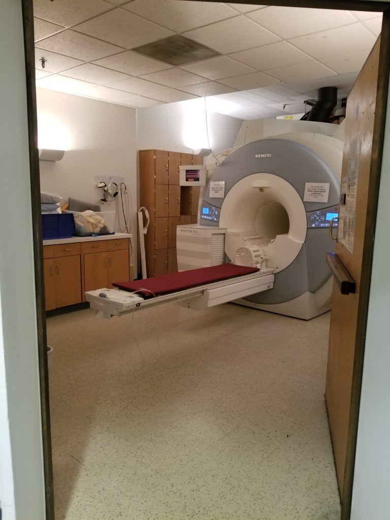
To meet these requirements, we use minimum norm MRI-constrained sourcespace reconstructions of simultaneously acquired MEG and EEG data. All neuromaging data are collected at the Athinoula A. Martinos Center for Biomedical Imaging using their Sieman’s 3T Trio and Prisma scanners, and the MEG facilities at the David Cohen MEG Laboratory, which include a 306 channel Neuromeg system with an integrated 70 channel EEG system all housed in a 6-layered Imelda magnetically shielded room. By leveraging anatomical and other constraints in the reconstruction process, we achieve source localization on roughly the scale of Brodmann units, and temporal resolution of 1 millisecond over all cortical surfaces. Our scanning team is led by Seppo Ahflors.


